The Foundations of Entrail Alignment Theory
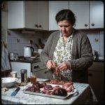
Entrail alignment theory holds that the diagnostic value of a reading lies not merely in the condition of individual organs, but in their spatial relationships to one another. A liver that would suggest caution in isolation may read quite differently when considered alongside a displaced spleen or an unusually positioned length of intestine. Most experienced practitioners already apply some version of this thinking intuitively. What a unified model attempts to do is make that intuition systematic — and therefore teachable, communicable, and subject to peer review.
The field has long suffered from a tendency towards organ-by-organ interpretation: the liver is examined, noted, set aside; the spleen follows; the intestines last. This sequential approach has its uses, particularly for newer practitioners still developing their vocabulary of form and colour. But it tends to underweight what is, in many readings, the most significant information available — the geometry of the whole.
The Core Variables of Alignment
A working model of entrail alignment must account for at least four classes of variable. These are not independent of one another, which is precisely why a unified approach is more useful than treating each in isolation.
Morphology: Shape and Scale
The physical form of each organ — its size relative to expectation, its surface texture, the regularity or irregularity of its margins — establishes the baseline from which relational readings are made. An enlarged organ occupies more positional space and may compress or displace adjacent structures, altering the alignment picture for the entire cavity. Practitioners who have read extensively on spleenfold mechanics will recognise that even modest variation in the spleen’s folding pattern can shift its apparent orientation relative to the stomach and left kidney, producing what superficially resembles a misalignment but is better understood as a morphological variant requiring adjusted interpretation.
Scale matters in a second sense as well. When working with smaller animals — rodents, poultry, rabbit — the organs are compressed into a much tighter spatial arrangement, reducing the interpretive margin between positions that would be clearly distinct in a larger specimen. This is one reason rodent haruspicy, while valuable, demands a level of positional precision that benefits from systematic training rather than intuition alone.
Spatial Relationships and Axial Orientation
The primary axis of a reading — broadly speaking, the cranio-caudal line from the diaphragm to the pelvic floor — provides the reference point from which lateral and dorsal displacement is measured. Organs that fall consistently to one side of this axis, or that present at an unexpected depth, are the first indicators that the alignment picture warrants careful attention.
Equally significant is the relationship between neighbouring organs: the proximity of the liver’s inferior margin to the transverse colon, the angle at which the gallbladder sits relative to the hepatic bed, the degree to which intestinal loops have settled into recognisable patterns versus presenting in apparent disorder. These relationships are not arbitrary. The body has preferred geometries, and deviation from them is part of what a reading records.
Environmental Influences
Temperature and humidity affect organ presentation more than many practitioners acknowledge in formal discussion, though most will quietly admit as much. Viscera presented in cold conditions contract and stiffen, compressing spatial relationships and making displacement harder to assess accurately. Warmer, more humid conditions produce the opposite effect — a certain looseness that can, if not accounted for, be misread as pathological displacement.
There is no universally agreed correction factor for environmental variables, which is one of the ongoing limitations of the field. What can be said is that consistent working conditions — same temperature range, same preparation interval, same surface and tray type — reduce this source of interpretive noise considerably. The guidance in storing organs safely at home is relevant here not only for hygiene reasons but for reading consistency.
Individual Variation
No two specimens present identically, and a unified model must be robust enough to accommodate genuine biological variation without collapsing into the position that anything goes. The practical solution most experienced practitioners arrive at is to maintain a working familiarity with population norms for the species most commonly used in their practice, so that individual variation can be distinguished from meaningful displacement.
This is one area where case records are invaluable. A practitioner who has documented fifty sheep readings has a calibrated internal reference that a practitioner who has done five simply does not. This is not a judgement — it is the ordinary reality of developing any perceptual skill.
Applying the Unified Model in Practice
The value of entrail alignment theory is not academic. It directly affects the reliability of readings, which affects the service offered to clients and, by extension, the reputation of the practice.
In practical terms, applying a unified model means resisting the urge to reach a conclusion before the full spatial picture has been assessed. It means noting not just what each organ looks like, but where it is — relative to the axis, relative to its neighbours, relative to expectation. It means developing a notation system, however informal, that allows a reading to be recorded with enough spatial information that it could be revisited or discussed with a colleague.
For practitioners working in autoharuspicy — a discipline that raises its own interpretive challenges, discussed more fully in our piece on unreliable organs — the unified model offers a structural anchor when subjective proximity to the material might otherwise distort the reading. Having a framework to return to is not a crutch; it is sound methodology.
Towards a Shared Framework
One of the longer-standing frustrations in haruspicy is the degree to which practitioners work in methodological isolation. Training routes vary enormously — as anyone who has spent time with a beginner’s guide to becoming a haruspex will have noted — and there is, as yet, no agreed standardisation of alignment terminology. A reading described as showing “lateral hepatic drift” by one practitioner may be recorded as “right-displaced liver” by another, referring to the same phenomenon.
This matters because shared language is a precondition of shared knowledge. The goal of a unified model is not to constrain individual practice or flatten the genuine interpretive differences that experience and tradition produce. It is to establish enough common ground that practitioners can compare findings, identify patterns across cases, and build a body of evidence that serves the whole field.
That ambition may seem premature given where the discipline currently stands institutionally. But the same ambition was present in every practice that eventually achieved a degree of professional coherence — and the work of building it begins with individual practitioners deciding that rigour is worth the effort, regardless of whether external recognition follows immediately.
Entrail alignment theory is, in that sense, less a finished model than an ongoing project. What matters is that we are working on it deliberately, and that the readings we perform today are better for the thinking we bring to them.
Subscribe to The Monthly Augury
A monthly reading of the entrails: our finest satire, curios and questionable prophecies, delivered once a moon.
Double opt-in — we'll email you one link to confirm. No spam, unsubscribe any time.
Equipment & References
 The Divine Liver — EllisonThe standard contemporary reference
 Veterinary Anatomy — Dyce et alHepatic and splenic morphology
 De Divinatione — CiceroFoundational text, ebook edition
As an Amazon Associate this site earns from qualifying purchases.